The Marco / Nidek OPD-Scan III instrument has brought waveform technology to Beartooth Vision Center. This instrument measures a wealth of information about the patient’s refractive status, including corneal topography (contour mapping of the clear tissue on the front of the eye), a person’s glare and blur simulations, and an aberrometer which measures the higher order aberrations (HOA’s) of the eye. For the first time in the United States, we have added the fitting software from the Waveform contact lens company to our OPD-Scan III to create a powerful problem-solving device for many of our patients who have special visual needs. The Waveform company manufactures highly precise custom wavefront guided soft contact lenses and multifocal contact lenses. With the fitting of Waveform contact lenses, Beartooth Vision Center has entered the cutting edge area of correction of higher order aberrations (HOA’s). There are about 80 different types of HOA’s. These are imperfections of light that occur as light goes through the pupil and different interfaces of the eye on the way to the retina in the back of the eye. Traditional glasses and contact lenses do not correct this blur that everyone has to one extent or the other.
The amount and impact of HOA’s vary widely from person to person. Some people have fewer HOA’s, with little effect on their vision. Other people have significant HOA – related visual problems, including glare and reduced clarity in low light and at night. People with degenerative corneal conditions (such as Keratoconus and Pellucid Marginal Degeneration) can experience significant distortion and blur of their vision under any lighting conditions – even with traditional glasses or contact lenses – due to an extreme amount of HOA’s from the corneal irregularities. With the incorporation of a precise “HOA fingerprint” custom soft contact lens correction compensated to align perfectly with the position and fit of the contact on the cornea, Waveform custom soft contacts can be life changing. These contact lenses are truly unique and custom made for our patients. Waveform contact lenses can provide far better visual clarity for these people, as well offering an alternative vision correction for people with very high prescriptions that create significant visual distortion with conventional lens technology. – Dr. Currence
Following is a quote from the Waveform web site:
 Over the past 15 years, new high-tolerance objective measuring technology called “wavefront aberrometry” has been developed. Conventional eye exams measure low order (2nd order) optical aberration—sphere, cylinder and axis and, in the case of a presbyope, the addition power. Through advancements in LASIK surgery, wavefront aberrometry was developed to optimize the visual outcome. Wavefront aberrometry is an advanced 21st century optical measuring technology that measures 28 levels of aberrations within the human eye, called Zernike coefficients. Only one of these levels—2nd order aberrations—are measured in conventional subjective refractions today. An aberrometer is capable of taking advanced, objective and complex optical measurements of the human eye in seconds, compared to the typical 15 minute subjective eye exam.
Optical aberrations are defined as anything that causes light to bend and can change significantly as the pupil dilates under low light conditions, such as at night. This is why many people are happy with their vision during daylight hours and then are uncomfortable and complain about their vision at night. The important optical aberrations that affect vision are:
- 2nd Order optical aberrations – currently measured in all eye exams providing sphere, cylinder and axis corrections
- 3rd and 4th Order optical aberrations – high order aberrations currently not measured in today’s eye exams but can account for up to 20% of the eye’s refractive error.
- 5th and 6th Order optical aberrations –also high order aberrations not currently measured in today’s eye exam. These aberrations are of less significance clinically, however they manifest in reduced vision for a small percentage of eyes.
Uncorrected high order aberrations (HOA’s) can lead to poor nighttime vision, a reduction in depth of focus for reading, glare around lights at night and reduced color perception. All of these HOA’s manifest in varying degrees for each individual patient. Pupil size is a significant contributor to the manifestation of high order aberrations. As the pupil increases in size, more peripheral light enters the eye. As a result, a blur circle is created instead of a point focus on the retina. As the blur circle increases in size and shape, visual clarity is diminished. This is why even some patients with 20/20 vision can complain of visual discomfort.
Wavefront aberrometry has been commercially available for over 10 years. Its primary use, when originally introduced into the optical industry, was to provide optical measurements that could aid the refractive surgeon improve the vision outcome for LASIK. The aberrometer shines a perfectly shaped wave of light into the eye and captures reflections distorted based on the eye’s surface contours. Thus, it generates a map of the optical system of the eye, which can be used to prescribe a solution, correcting the patient’s specific vision problem.
Over the past four or five years, this technology has greatly improved optometrists and ophthalmologists’ ability to develop more precise and specific prescriptions. By using the wavefront aberrometer, they have helped many patients achieve a much higher standard of vision.
WaveForm has developed two technologies that utilize advanced wavefront aberrometry measurements to improve the eye exam experience called “Optimized WaveFont Refraction,” the patient and eye doctor can provide a customized correction of each individual’s low and high order optical aberrations through the manufacture of wavefront-guided soft contact lenses.
WaveForm’s wavefront-guided contact lens technology and optimized wavefront refraction for eyeglass lenses address the problem of uncorrected high order aberrations in different ways.
- For WaveForm’s wavefront-based objective eye exams, the optimized wavefront refraction incorporates all low and high order aberrations to produce an optimal low order prescription in seconds. Since the eye moves behind the eyeglass lens, it is virtually impossible to correct for the optical fingerprint on the surface of eyeglass lenses.
- In the case of WaveForm’s wavefront-guided contact lens technology, the individual optical fingerprint is generated on the surface of the soft contact lenses and placed directly on the eye’s surface, thereby fully correcting this unique optical fingerprint.

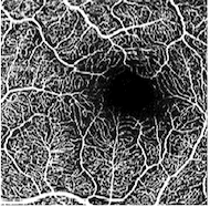
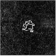
 There are no other instruments like the MonacoPro and its predecessor, the Daytona scanning retinal laser. We have used it as an important part of our annual eye examinations since 1998. The MonacoPro retinal exam is an option our patients can choose as part of their yearly eye exam to evaluate the health of the inside of their eyes. It gives a digital ultra-widefield view of the internal eye, including the retina, the optic nerve, the blood vessels, and the macula (the part of the retina that provides your sharp central vision). This technology has revealed diabetes, cancer, high blood pressure, blockage of the blood vessels to the eye, glaucoma, macular degeneration, retinal detachments, tears, and holes, as well as other retinal conditions in our patient’s eyes. It has been a sight-saver for many of our patients over the years.
 We acquired our Heidelberg Spectralis Ocular Coherence Tomographer (OCT) in October, 2010. An OCT is essentially a high resolution CATSCAN of the retina, the cornea (the clear tissue on the front of the eye) and other internal ocular features. The OCT uses a series of very detailed and highly magnified visual slices through the tissue in question that allows us to closely evaluate problem areas we discover inside the eye using the Optomap instrument (see above).

 Our Humphrey visual field analyser is the gold standard for measuring the peripheral visual field. In healthy eyes, the peripheral vision (also known as “side vision”) remains the same in size throughout life. There are medical conditions such as glaucoma, stroke, and space – occupying lesions inside the brain which can cause complete or partial loss of sensitivity of the peripheral vision called an absolute (complete) or relative (partial) scotomas. We have used both of these instruments to detect significant medical problems and both will continue to be valuable diagnostic tools at Beartooth Vision Center. – Dr. Currence
 The addition of the Reichert Ocular Response Analyzer G3 provides our office the ability to understand even better the nuances and dynamics of glaucoma. Corneal hysteresis is a measure of the behavior of the eye in that it measures how a cornea responds to an outside pressure. It is essentially a measure of the elasticity of an eye. Some eyes are very elastic (like a balloon) and can absorb a higher internal pressure with no damage. Other eyes are far more rigid (like a glass bottle) and are much more likely to have damage with a higher pressure inside. The Reichert Ocular Response Analyzer G3 is a very good predictor of who is at risk of developing glaucoma and also how well our glaucoma patients are responding to their medications. – Dr. Currence